

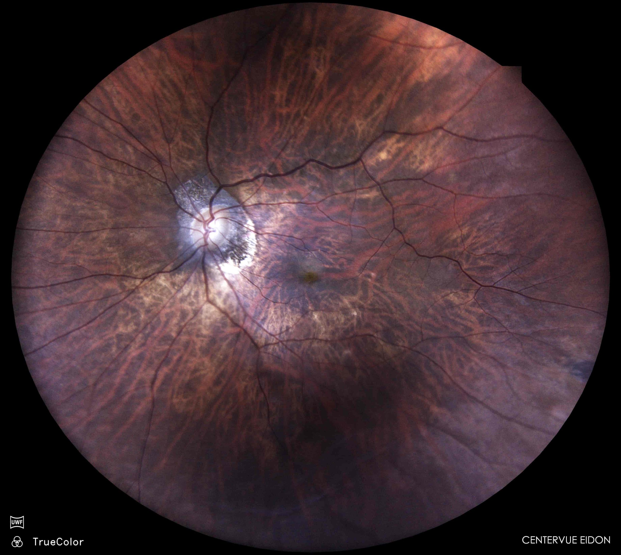
Description:Myoipc CNVM is a vision-threatening complication that often remains clinically subtle due to background atrophic changes. FFA helps in diagnosis by revealing classic CNVM seen as early well defined hyperfluorescence with late leakage.Lacquer cracks are known precursors of myopic CNV. Early identification is essential as Myopic CNVM responds to anti-VEGF therapy.

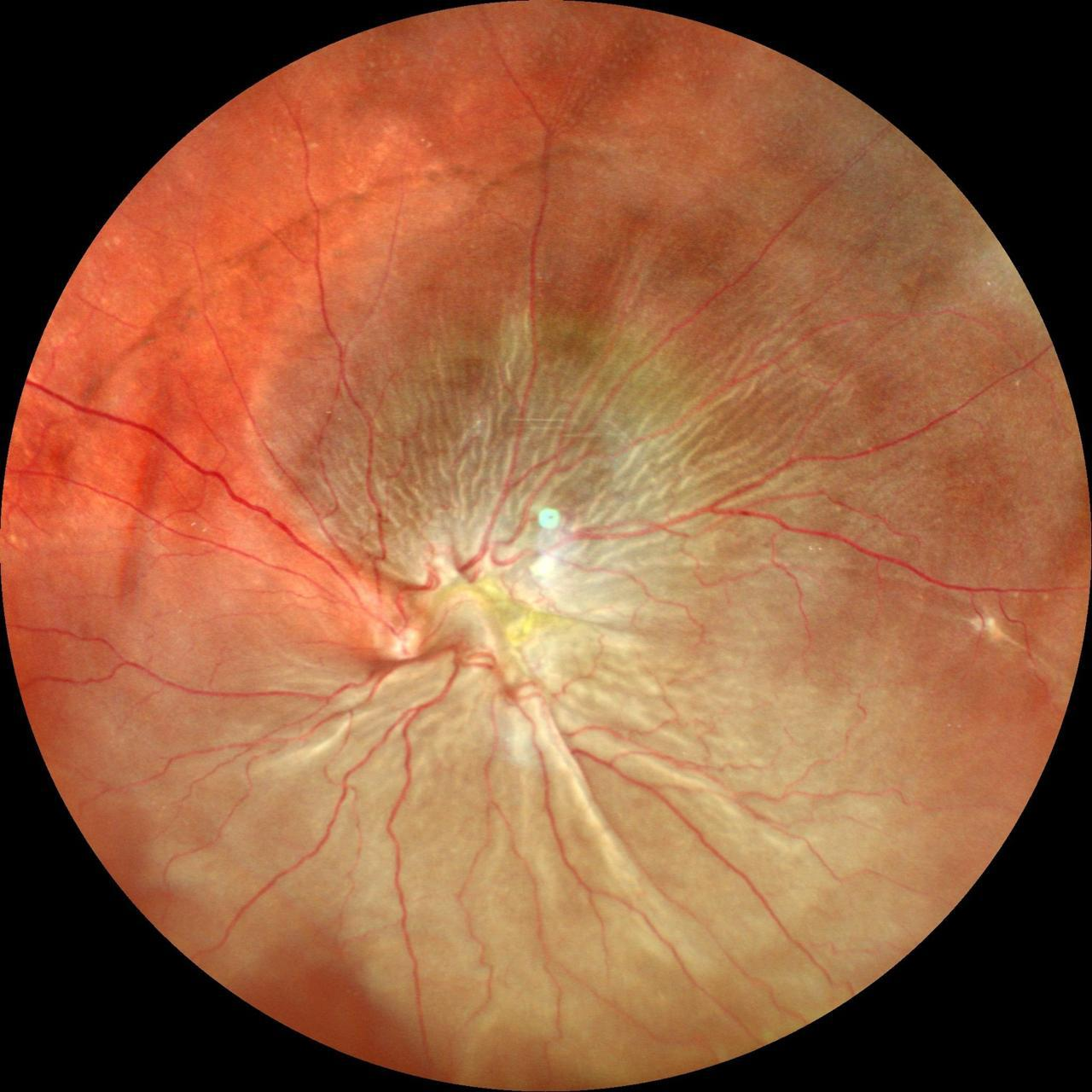
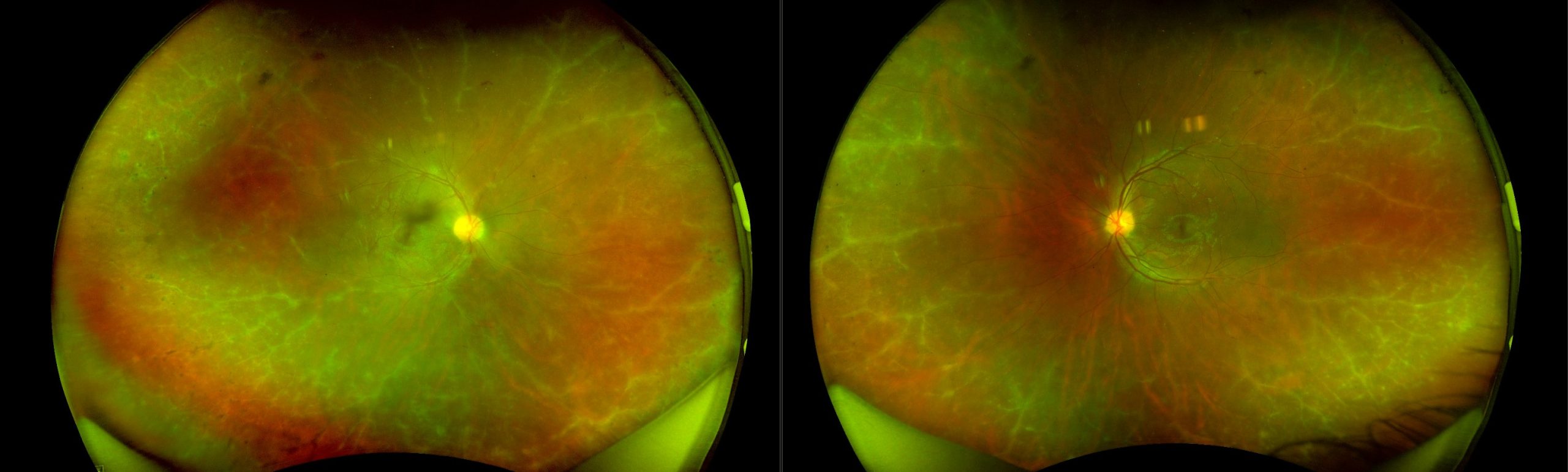
Description: “Fundus photography of the left eye reveals a subtotal retinal detachment with significant macular involvement, complicated by proliferative vitreoretinopathy (PVR). The macula exhibits a characteristic ‘crumpled’ appearance, indicative of advanced PVR and retinal contraction.”

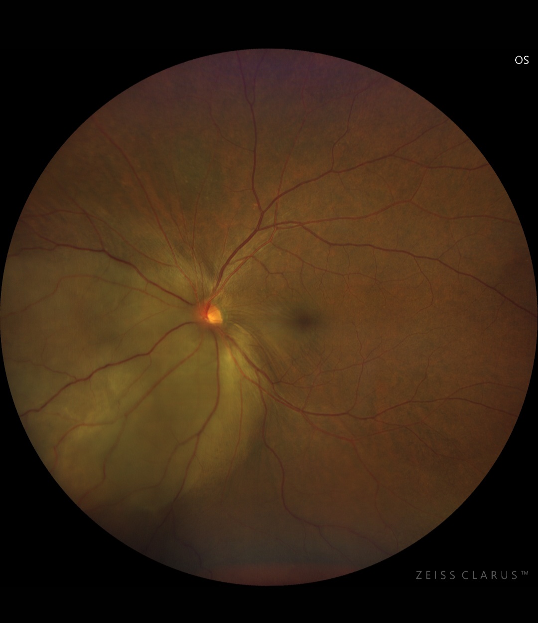
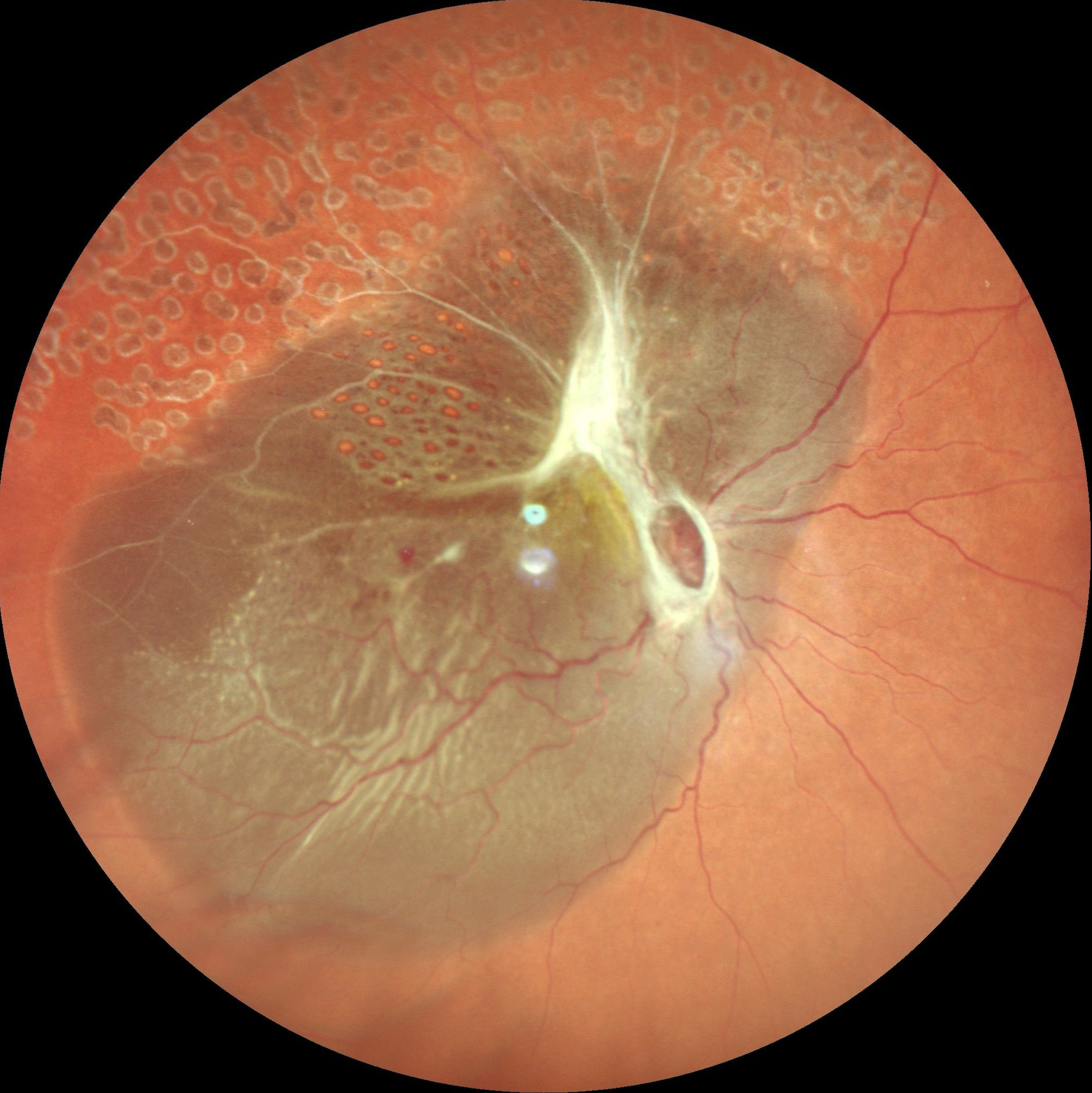






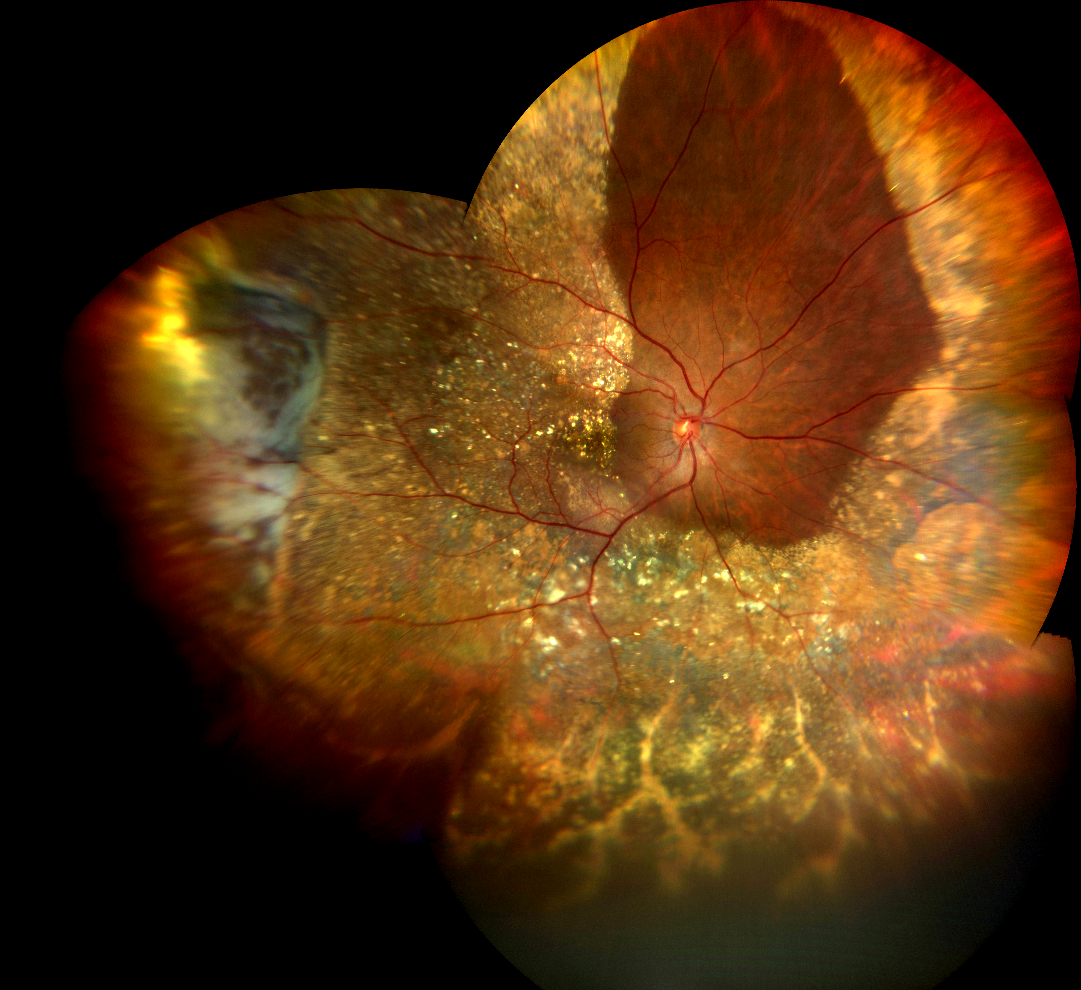
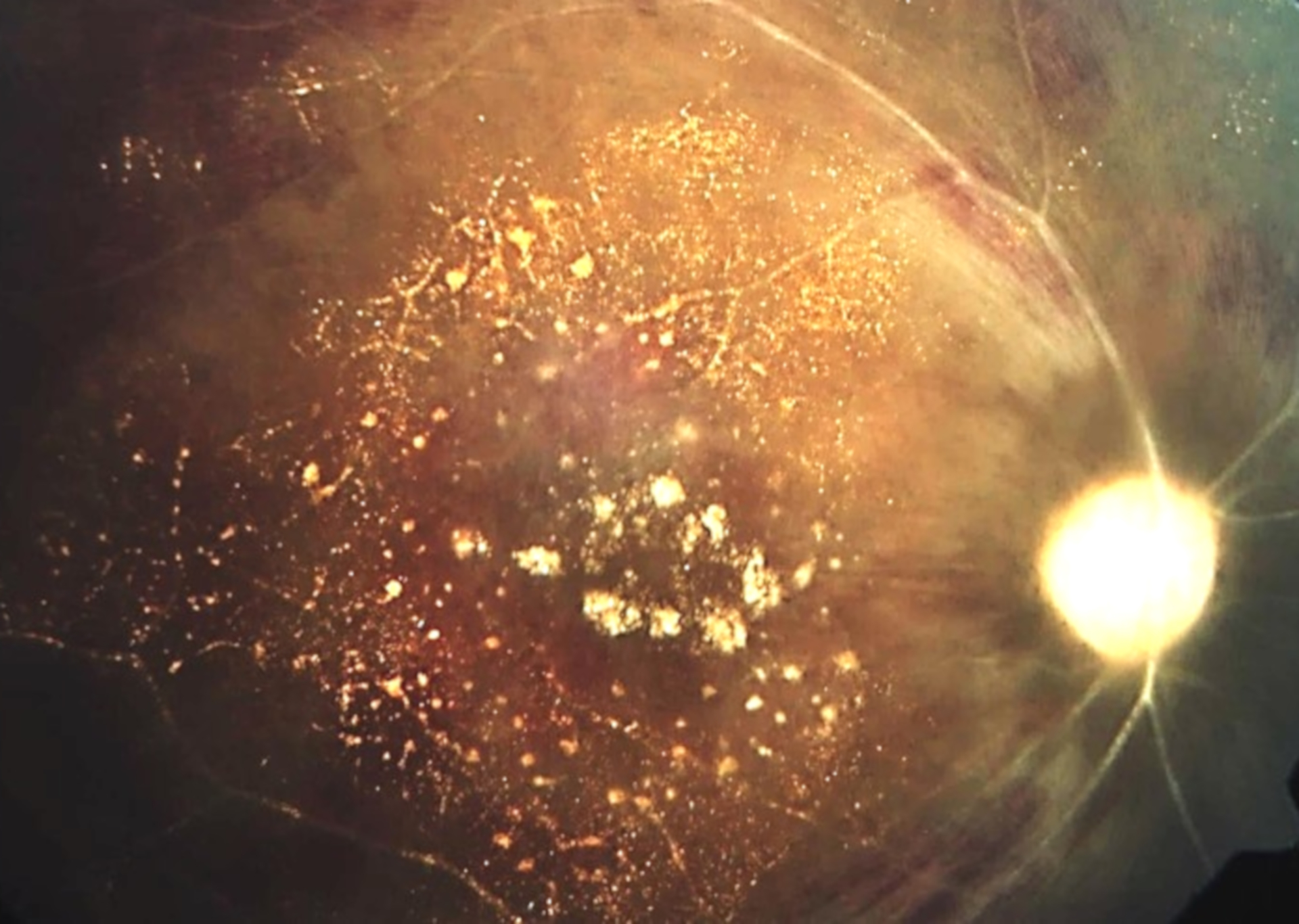
Description: The image shows a post vitrectomised eye with crystalline subretinal deposits


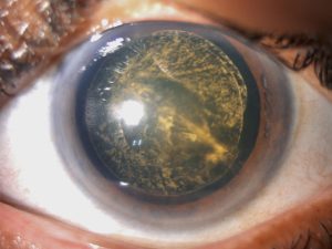
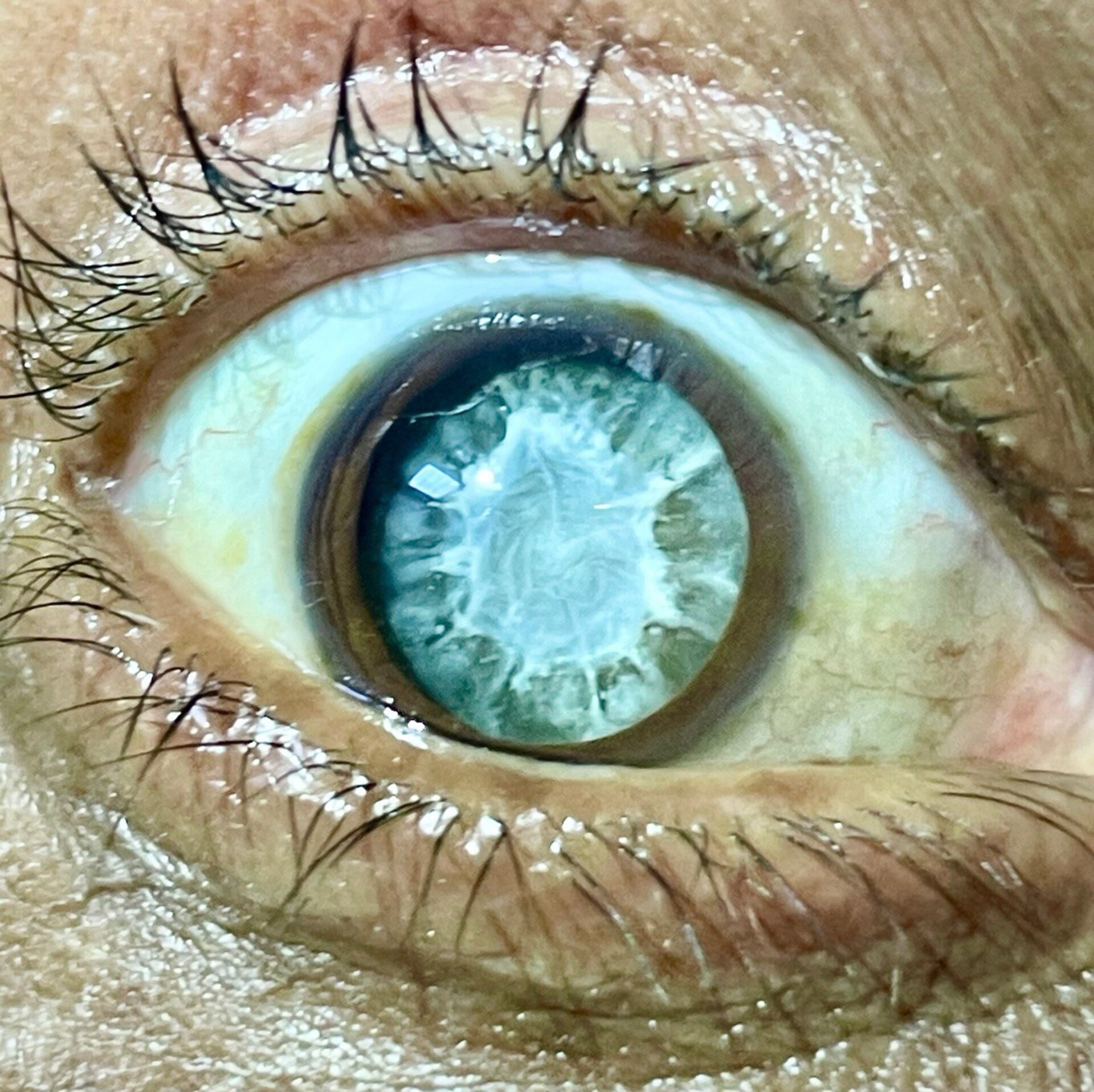
Description: This image illustrates an accidental intralenticular injection of intravitreal triamcinolone acetonide (IVTA), characterized by the deposition of IVTA crystals within the lens and a clearly visible needle track. The patient was successfully managed with phacoemulsification and intraocular lens (IOL) implantation.

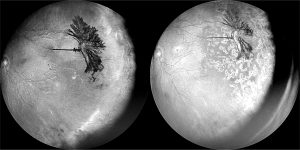
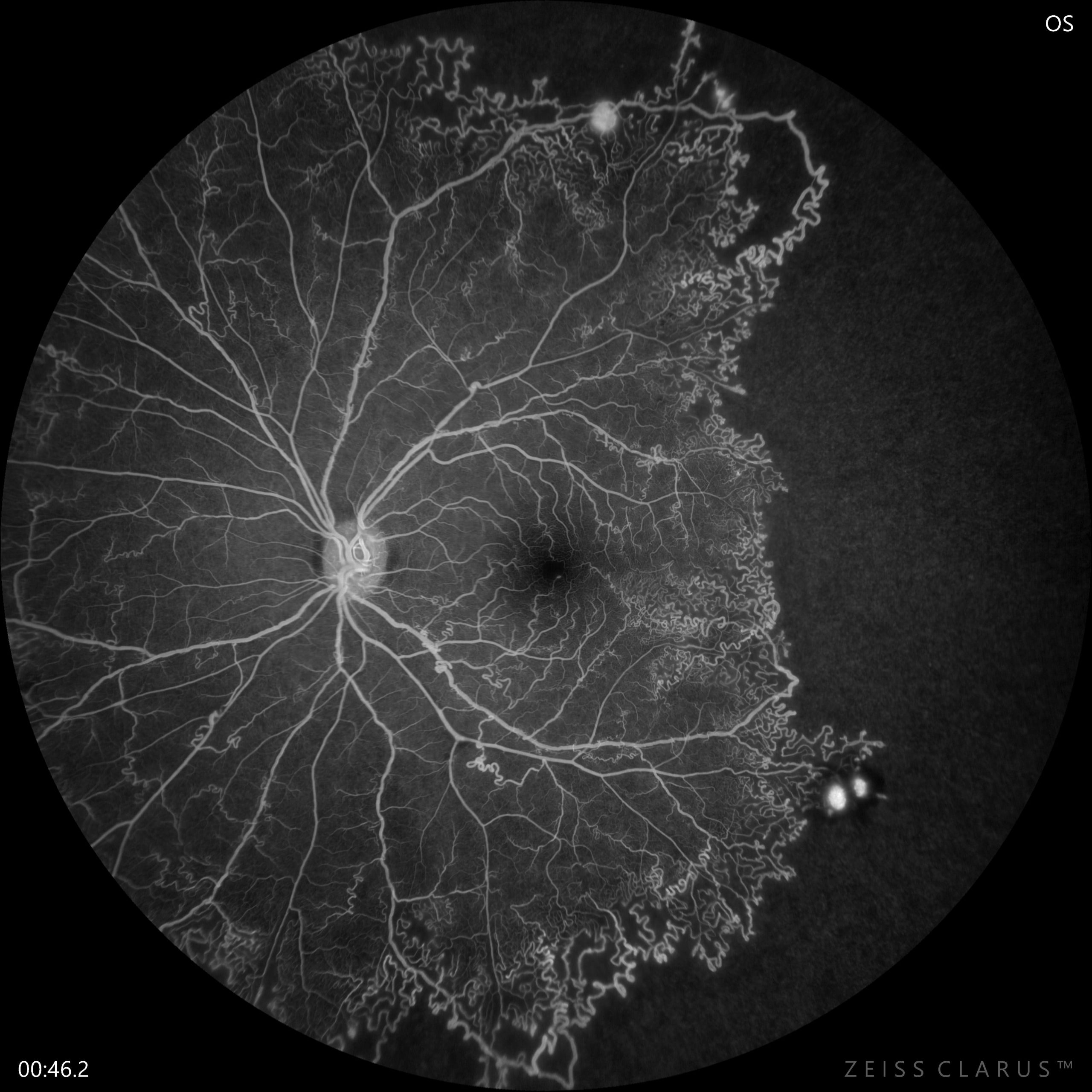
Description: These are Monochromatic images of the left eye of a patient with Systemic Lupus Erythematosus (SLE) displaying distinct ocular manifestations. The first image, on the left, captures sea fan neovascularization at its peak, flourishing over an ischemic retina. The second image, on the right, reveals partial regression of the neovascularization, six weeks after undergoing targeted laser photocoagulation. The progression is evocative of a tree in full bloom during spring, gradually withering away as autumn approaches.